韓国美容外科アカデミー フェイスリフト 韓国 ソウル 2009/06/13-14
招待講演: facelift video text
video presentation of facelift “treatment of igament, SMAS and fat tissue in facelift surgery”
■ 01
(01)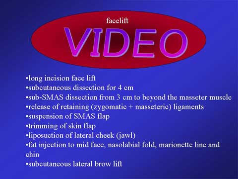
・This video presentation demonstrates the facelift procedure on a female patient who presented with jawl deformity, deep nasolabial fold and marionette line, volume loss in midface, submental fat deposit and upper eyelid hooding. The operation consists of long incision face lift
・subcutaneous dissection for 4 cm
・sub-SMAS dissection from 3 cm to beyond the masseter muscle
・elease of retaining (zygomatic + masseteric) ligaments
・suspension of SMAS flap
・trimming of skin flap
・liposuction of lateral cheek (jawl)
・fat injection to mid face, nasolabial fold, marionette line and chin
・subcutaneous lateral brow lift
■ 02
(02)
The retaining ligaments suspend the facial skin to the underlying deep structure. As aging, the soft tissue (skin and subcutaneous fat) looses the firmness. The skin between the masseteric retaining ligament and mandibular retaining ligament show the sag over the mandibular ligaments due to gravity in the upright position. This sagging deformity is recognized as jawl.
■ 03
(03)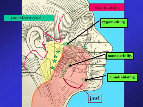
A long skin incision is made along the temporal hairline, the side burn, natural crease in front of the ear, the ear lobe, postauricular groove and occipital hairline. The lateral margin of platysma and muscular portion of SMAS is located at 3 cm from the ear lobe.
■ 04
(04)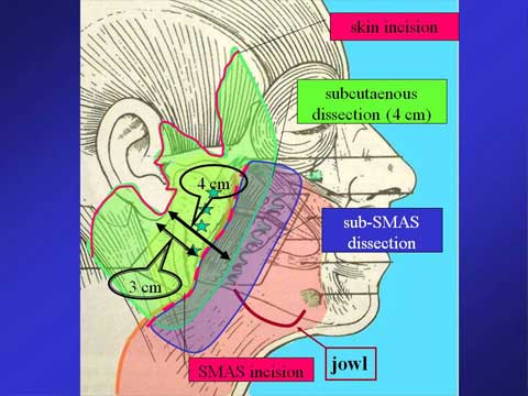
The subcutaneous dissection is made up to 4 cm from the ear lobe. The SMAS is incised at the lateral margin of the platysma which is at 3 cm from the ear lobe. The deep dissection is carried out under the SMAS and platysma and continued up to beyond the anterior margin of the masseteric muscle where the masseteric ligaments are present. This sub SMAS dissection can release all the masseteric ligament s and zygomatic ligaments from the SMAS, which allow us to pull the medial portion of the skin and SMAS with traction of the SMAS flap.
■ 05
(05)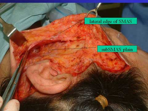
The intraoperative photo showing the 1cm long SMAS cuff attached to the skin flap and wide sub-SMAS pocket.
■ 06
(06)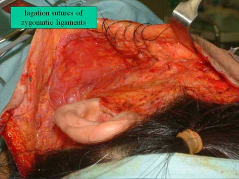
The suspension of the short SMAS flap to the mastoid fascia and relatively immobile SMAS over the parotid gland.
■ 07
(07)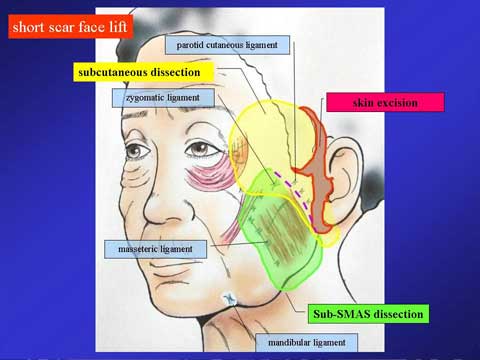
In case of no significant neck sagging, the facelift with a short scar is feasible. This procedure eliminates an incision along the occipital hairline. Instead of using the temporal hairline incision, an incision is made in the hair-baring skin the temporal area.
■ 08
(08)
A 42 year old female presented with mild jawl deformity and heavy look in the upper eyelid.
■ 09
(Figure 09)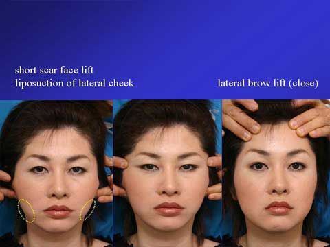
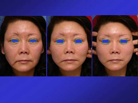
For her preoperative planning, traction of skin in front of the ear with fingers demonstrates the possible result from the short scar facelift. The simulation shows budging in the lateral cheek in spite of the strong pull. In order to make smaller the bulge which will remain after facelift alone, the combination of liposuction with facelift is useful. The middle picture shows the simulation of facelift with temporal lift, which make the lateral canthus look sharper. The patient denied to have temporal lift. The right picture demonstrates possible result from lateral brow lift.
■ 10
(10)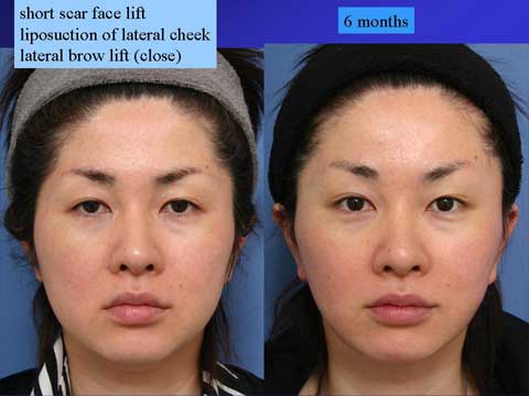
6 months after the short scar facelift with liposuction in the lateral cheek and lateral brow lift with close approach.
■ 11
(11)
62 year-old female presented with jawl, marionette line, nasolabial fold and loss of soft tissue fullness. The middle picture shows simulation of facelift for the lateral cheek. The right picture shows simulation of facelift and temporal lift which provide the lateral canthal region and lower eyelid with pull in the super-lateral direction. The patient chose to have temporal lift with pulled appearance of the lateral canthus.
■ 12
(12)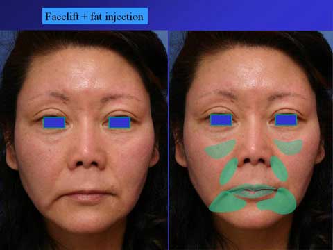
The green marking demonstrates the area for which lipofilling is planned.
■ 13
(13)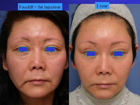
One year after facelift and temporal lift with lipo-injection.
Live surgery
フェイスリフトのデモ手術の患者さんに手術のデザインをして、今から手術。
(写真A)
(写真A)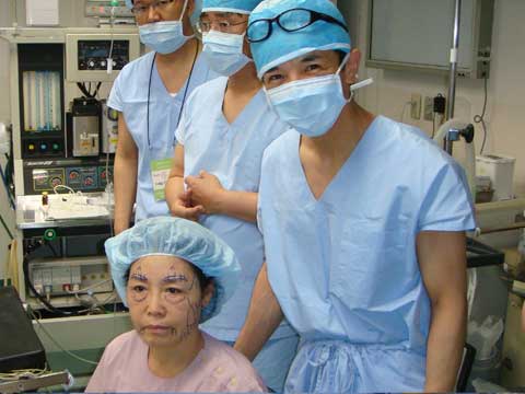
フェイスリフトのライブサージェリーのデモ中。(写真B)
(写真B)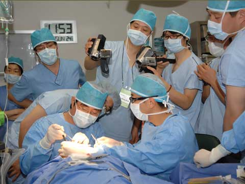
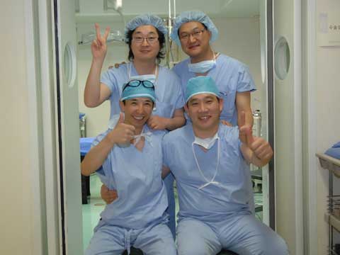
手術が終了。助手をしてくれた韓国の先生方と。(写真C)
(写真C)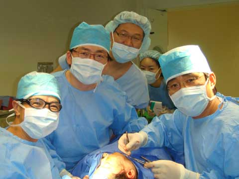
手術が無事終了して、助手をしてくれた先生とアカデミーの会長キム先生と記念撮影。(写真D)
(写真D)